Electrophysiology
Electrophysiology (EP) Cardiology
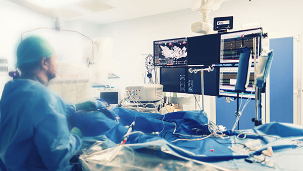
Electrophysiology (EP) testing involves placing a special catheter into the right side of the heart that relates to the electrical pathway or conduit of the heart. The electrical pathways are mapped out by the electrophysiologist to see where the irregular heart rhythms arise. This allows the physician to ablate (burn) at a precise location to re-direct and restore a patient’s normal heart rhythm.
What is Electrophysiology Cardiology?
It's the study of the heart's electrical system. The term "EP study" applies to any
procedure that includes the insertion of an electrode catheter into the heart. Electrode catheters are long, flexible wires that transmit electrical currents to and from the heart. Some EP studies are done to diagnose abnormalities, while others are done to access the heart for treatment of the problem.
procedure that includes the insertion of an electrode catheter into the heart. Electrode catheters are long, flexible wires that transmit electrical currents to and from the heart. Some EP studies are done to diagnose abnormalities, while others are done to access the heart for treatment of the problem.
Common EP Procedures include:
- Diagnostic EP study
- Cardiac Ablation
- An internal cardiac defibrillator can perform the same functions as a permanent pacemaker. The difference with this device is it has the capability to send a small electrical shock to the heart if a deadly heart rhythm is detected.
- A pacemaker is implant when a heart is not beating fast enough or has demonstrated abnormal electrical sign which cause you heart to beat irregular. A permanent pacemaker is implanted by creating a small incision in the upper chest wall and has a 7 to 10-year life.
- Cardioversion
Someone who has an abnormal heart rhythm – a heart that beats too slowly, too rapidly, or in an irregular pattern would be a good candidate for an EP procedure. Having an irregular heart rhythm diminishes the pumping power of the heart and results in poor circulation. When the body is not getting the oxygen it needs, a person experiences shortness of breath, dizziness, palpitations, and other symptoms. From catheter ablations to working with patients who have pacemakers and defibrillators, an Electrophysiologist works as an “electrician of the heart,” to correct hearts that beat too slow or too fast.
A Cardioversion is the use of electric current to “shock” your heart back into a normal rhythm. For this procedure you will be given medication to make you sleep. Before the cardioversion you will need a special ultrasound called a Transesophageal Echocardiogram (TEE). This is an ultrasound of your heart obtained through your throat (esophagus). A small tube with a camera on the end of it will pass through your throat to take ultrasound pictures of the backside of your heart. The pictures show more detail than those obtained from the surface of your chest. This will help your doctor make sure there are no blood clots in your heart before your cardioversion.

